- Science Notes Posts
- Contact Science Notes
- Todd Helmenstine Biography
- Anne Helmenstine Biography
- Free Printable Periodic Tables (PDF and PNG)
- Periodic Table Wallpapers
- Interactive Periodic Table
- Periodic Table Posters
- How to Grow Crystals
- Chemistry Projects
- Fire and Flames Projects
- Holiday Science
- Chemistry Problems With Answers
- Physics Problems
- Unit Conversion Example Problems
- Chemistry Worksheets
- Biology Worksheets
- Periodic Table Worksheets
- Physical Science Worksheets
- Science Lab Worksheets
- My Amazon Books

Human Anatomy Worksheets and Study Guides

This is a collection of free human anatomy worksheets. The completed worksheets make great study guides for learning bones, muscles, organ systems, etc. The worksheets come in a variety of formats for downloading and printing. In most cases, the PDF worksheets print the best. But, you may prefer to work online with Google Slides or print the PNG images.
Do you need a particular worksheet, but don’t see it? Ideas for worksheet topics you want covered are welcome!
Human Anatomy Worksheets
These worksheets cover major organs and organ systems.

Label the Heart
Label the parts of the human heart.
[ Google Apps worksheet ][ worksheet PDF ][ worksheet PNG ][ answers PNG ]

Label the Eye
Label the parts of the eye.
[ Google Apps worksheet ][ worksheet PDF ][ answers PDF ][ worksheet PNG ]

Types of Blood Cells
Identify the types of blood cells.
[ worksheet Google Apps ][ worksheet PDF ][ worksheet PNG ][ answers PNG ]

Label the Muscles
Label the major anterior muscles.
[ worksheet PDF ][ worksheet PNG ][ answers PNG ]

Label the Ear
Label the human ear.
[ Google Apps worksheet ][ Worksheet PDF ][ Worksheet PNG ][ Answers PNG ]

Label the Lungs
Identify the parts of the lungs.

Label the Kidney
Label the parts of the kidney.

Label the Liver
Identify the anatomy of the liver.

Label the Large Intestine
Label the parts of the large intestine.

Label the Stomach
Label the human stomach.
[ Google Apps worksheet ] [Worksheet PDF ][ Worksheet PNG ][ Answers PNG ]

External Nose Anatomy
Identify the parts of the nose.
[ Worksheet PDF ][ Worksheet Google Apps ][ Worksheet PNG ][ Answers PNG ]

Parts of the Nose
Here’s another way of identifying nose anatomy.

Label Bones of the Skeleton
Identify major bones of the skeleton.
[ Google Apps worksheet ][ worksheet PDF ][ answers PDF ][ worksheet PNG ][ answers PNG ]

Label the Lymph Node
Label the lymph node.

Label the Human Skull
[ worksheet PDF ][ worksheet Google Apps ][ worksheet PNG ][ answers PNG ]

Label the Skull (Advanced)

Label the Parts of the Brain
Identify parts of a human brain.

Label the Lobes of the Brain
Identify the different lobes of the brain.

Brain Anatomical Sections
Explore anatomical sections using a human brain as a reference.

Arteries of the Brain
Identify major brain arteries.

Label the Pancreas
Label the parts of the human pancreas.

Label the Spleen
Label spleen anatomy.

Label the Digestive System
Identify parts of the human digestive system.

Label the Respiratory System
Label the respiratory system.

Parts of a Neuron
Identify parts of a neuron.

Label the Lips
Label human lips.

Label the Skin
Label layers and structures in skin.

Label the Circulatory System
Label the circulatory system.

The Urinary Tract
[ Worksheet PDF ][ Worksheet Google Apps ][ Worksheet PNG ][ Answer Key PNG ]

The Bladder

The Female Reproductive System

Label Human Teeth

Identify Organs #1

Identify Organ Systems #1

Identify Organs #2

Identify Organ Systems #2
- Diagram of the Human Eye [ JPG ]
Human Anatomy Worksheets Terms of Use
You are welcome to print these resources for personal or classroom use. They may be used as handouts or posters. They may not be posted elsewhere online, sold, or used on products for sale.
This page doesn’t include all of the assets on the Science Notes site. If there’s a table or worksheet you need but don’t see, just let us know. The same goes if you need a different file format.
Related Posts

1.4 Anatomical Terminology
Learning objectives.
By the end of this section, you will be able to:
- Use appropriate anatomical terminology to identify key body structures, body regions, and directions in the body
- Demonstrate the anatomical position
- Describe the human body using directional and regional terms
- Identify three planes most commonly used in the study of anatomy
- Distinguish between major body cavities
Anatomists and health care providers use terminology that can be bewildering to the uninitiated; however, the purpose of this language is not to confuse, but rather to increase precision and reduce medical errors. For example, is a scar “above the wrist” located on the forearm two or three inches away from the hand? Or is it at the base of the hand? Is it on the palm-side or back-side? By using precise anatomical terminology, we eliminate ambiguity. For example, you might say a scar “on the anterior antebrachium 3 inches proximal to the carpus”. Anatomical terms are derived from ancient Greek and Latin words. Because these languages are no longer used in everyday conversation, the meaning of their words do not change.
Anatomical terms are made up of roots, prefixes, and suffixes. The root of a term often refers to an organ, tissue, or condition, whereas the prefix or suffix often describes the root. For example, in the disorder hypertension, the prefix “hyper-” means “high” or “over,” and the root word “tension” refers to pressure, so the word “hypertension” refers to abnormally high blood pressure.
Anatomical Position
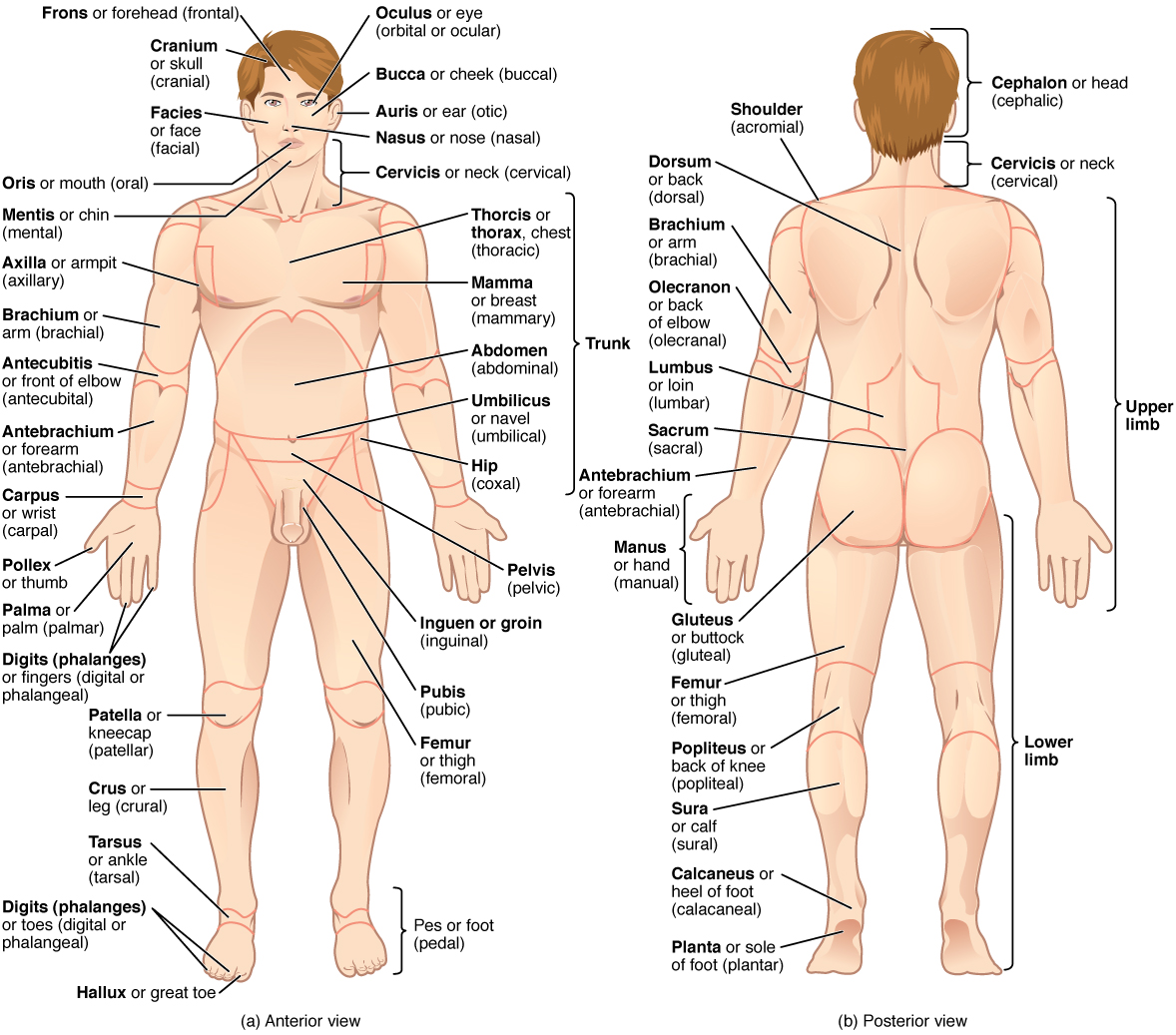
To further increase precision, anatomists standardize the way in which they view the body. Just as maps are normally oriented with north at the top, the standard body “map,” or anatomical position , is that of the body standing upright, with the feet at shoulder width and parallel, toes forward. The upper limbs are held out to each side, and the palms of the hands face forward as illustrated in Figure 1.4.1 . Using this standard position reduces confusion. It does not matter how the body being described is oriented, the terms are used as if it is in anatomical position. For example, a scar in the “anterior (front) carpal (wrist) region” would be present on the palm side of the wrist. The term “anterior” would be used even if the hand were palm down on a table.

A body that is lying down is described as either prone or supine. Prone describes a face-down orientation, and supine describes a face up orientation. These terms are sometimes used in describing the position of the body during specific physical examinations or surgical procedures.
Regional Terms
The human body’s numerous regions have specific terms to help increase precision (see Figure 1.4.1 ). Notice that the term “brachium” or “arm” is reserved for the “upper arm” and “antebrachium” or “forearm” is used rather than “lower arm.” Similarly, “femur” or “thigh” is correct, and “leg” or “crus” is reserved for the portion of the lower limb between the knee and the ankle. You will be able to describe the body’s regions using the terms from the figure.
Directional Terms
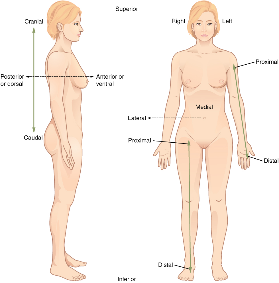
Certain directional anatomical terms appear throughout this and any other anatomy textbook ( Figure 1.4.2 ). These terms are essential for describing the relative locations of different body structures. For instance, an anatomist might describe one band of tissue as “inferior to” another or a physician might describe a tumor as “superficial to” a deeper body structure. Commit these terms to memory to avoid confusion when you are studying or describing the locations of particular body parts.
- Anterior (or ventral ) describes the front or direction toward the front of the body. The toes are anterior to the foot.
- Posterior (or dorsal ) describes the back or direction toward the back of the body. The popliteus is posterior to the patella.
- Superior (or cranial ) describes a position above or higher than another part of the body proper. The orbits are superior to the oris.
- Inferior (or caudal ) describes a position below or lower than another part of the body proper; near or toward the tail (in humans, the coccyx, or lowest part of the spinal column). The pelvis is inferior to the abdomen.
- Lateral describes the side or direction toward the side of the body. The thumb (pollex) is lateral to the digits.
- Medial describes the middle or direction toward the middle of the body. The hallux is the medial toe.
- Proximal describes a position in a limb that is nearer to the point of attachment or the trunk of the body. The brachium is proximal to the antebrachium.
- Distal describes a position in a limb that is farther from the point of attachment or the trunk of the body. The crus is distal to the femur.
- Superficial describes a position closer to the surface of the body. The skin is superficial to the bones.
- Deep describes a position farther from the surface of the body. The brain is deep to the skull.

Body Planes
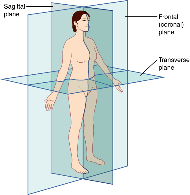
A section is a two-dimensional surface of a three-dimensional structure that has been cut. Modern medical imaging devices enable clinicians to obtain “virtual sections” of living bodies. We call these scans. Body sections and scans can be correctly interpreted, only if the viewer understands the plane along which the section was made. A plane is an imaginary, two-dimensional surface that passes through the body. There are three planes commonly referred to in anatomy and medicine, as illustrated in Figure 1.4.3 .
- The sagittal plane divides the body or an organ vertically into right and left sides. If this vertical plane runs directly down the middle of the body, it is called the midsagittal or median plane. If it divides the body into unequal right and left sides, it is called a parasagittal plane or less commonly a longitudinal section.
- The frontal plane divides the body or an organ into an anterior (front) portion and a posterior (rear) portion. The frontal plane is often referred to as a coronal plane. (“Corona” is Latin for “crown.”)
- The transverse (or horizontal) plane divides the body or organ horizontally into upper and lower portions. Transverse planes produce images referred to as cross sections.

Body Cavities
The body maintains its internal organization by means of membranes, sheaths, and other structures that separate compartments. The main cavities of the body include the cranial, thoracic and abdominopelvic (also known as the peritoneal) cavities. The cranial bones create the cranial cavity where the brain sits. The thoracic cavity is enclosed by the rib cage and contains the lungs and the heart, which is located in the mediastinum. The diaphragm forms the floor of the thoracic cavity and separates it from the more inferior abdominopelvic/peritoneal cavity. The abdominopelvic/peritoneal cavity is the largest cavity in the body. Although no membrane physically divides the abdominopelvic cavity, it can be useful to distinguish between the abdominal cavity, (the division that houses the digestive organs), and the pelvic cavity, (the division that houses the organs of reproduction).
Abdominal Regions and Quadrants
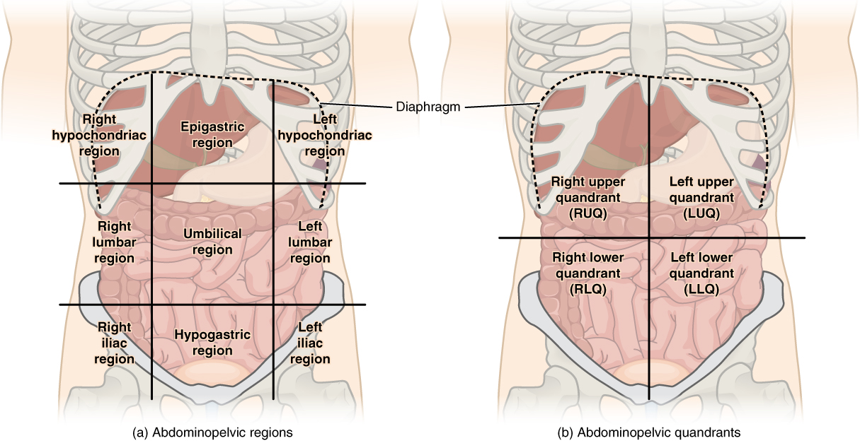
To promote clear communication, for instance, about the location of a patient’s abdominal pain or a suspicious mass, health care providers typically divide up the cavity into either nine regions or four quadrants ( Figure 1.4.4 ).

The more detailed regional approach subdivides the cavity with one horizontal line immediately inferior to the ribs and one immediately superior to the pelvis, and two vertical lines drawn as if dropped from the midpoint of each clavicle (collarbone). There are nine resulting regions. The simpler quadrants approach, which is more commonly used in medicine, subdivides the cavity with one horizontal and one vertical line that intersect at the patient’s umbilicus (navel).
Chapter Review
Ancient Greek and Latin words are used to build anatomical terms. A standard reference position for mapping the body’s structures is the normal anatomical position. Regions of the body are identified using terms such as “occipital” that are more precise than common words and phrases such as “the back of the head.” Directional terms such as anterior and posterior are essential for accurately describing the relative locations of body structures. Images of the body’s interior commonly align along one of three planes: the sagittal, frontal, or transverse.
Review Questions
Critical thinking questions.
In which direction would an MRI scanner move to produce sequential images of the body in the frontal plane, and in which direction would an MRI scanner move to produce sequential images of the body in the sagittal plane?
If the body were supine or prone, the MRI scanner would move from top to bottom to produce frontal sections, which would divide the body into anterior and posterior portions, as in “cutting” a deck of cards. Again, if the body were supine or prone, to produce sagittal sections, the scanner would move from left to right or from right to left to divide the body lengthwise into left and right portions.
This work, Anatomy & Physiology, is adapted from Anatomy & Physiology by OpenStax , licensed under CC BY . This edition, with revised content and artwork, is licensed under CC BY-SA except where otherwise noted.
Images, from Anatomy & Physiology by OpenStax , are licensed under CC BY except where otherwise noted.
Access the original for free at https://openstax.org/books/anatomy-and-physiology/pages/1-introduction .
Anatomy & Physiology Copyright © 2019 by Lindsay M. Biga, Staci Bronson, Sierra Dawson, Amy Harwell, Robin Hopkins, Joel Kaufmann, Mike LeMaster, Philip Matern, Katie Morrison-Graham, Kristen Oja, Devon Quick, Jon Runyeon, OSU OERU, and OpenStax is licensed under a Creative Commons Attribution-ShareAlike 4.0 International License , except where otherwise noted.

Introduction
Chapter objectives.
After studying this chapter, you will be able to:
- Distinguish between anatomy and physiology, and identify several branches of each
- Describe the structure of the body, from simplest to most complex, in terms of the six levels of organization
- Identify the functional characteristics of human life
- Identify the four requirements for human survival
- Define homeostasis and explain its importance to normal human functioning
- Use appropriate anatomical terminology to identify key body structures, body regions, and directions in the body
- Compare and contrast at least four medical imaging techniques in terms of their function and use in medicine
Though you may approach a course in anatomy and physiology strictly as a requirement for your field of study, the knowledge you gain in this course will serve you well in many aspects of your life. An understanding of anatomy and physiology is not only fundamental to any career in the health professions, but it can also benefit your own health. Familiarity with the human body can help you make healthful choices and prompt you to take appropriate action when signs of illness arise. Your knowledge in this field will help you understand news about nutrition, medications, medical devices, and procedures and help you understand genetic or infectious diseases. At some point, everyone will have a problem with some aspect of their body and your knowledge can help you to be a better parent, spouse, partner, friend, colleague, or caregiver.
This chapter begins with an overview of anatomy and physiology and a preview of the body regions and functions. It then covers the characteristics of life and how the body works to maintain stable conditions. It introduces a set of standard terms for body structures and for planes and positions in the body that will serve as a foundation for more comprehensive information covered later in the text. It ends with examples of medical imaging used to see inside the living body.
As an Amazon Associate we earn from qualifying purchases.
This book may not be used in the training of large language models or otherwise be ingested into large language models or generative AI offerings without OpenStax's permission.
Want to cite, share, or modify this book? This book uses the Creative Commons Attribution License and you must attribute OpenStax.
Access for free at https://openstax.org/books/anatomy-and-physiology-2e/pages/1-introduction
- Authors: J. Gordon Betts, Kelly A. Young, James A. Wise, Eddie Johnson, Brandon Poe, Dean H. Kruse, Oksana Korol, Jody E. Johnson, Mark Womble, Peter DeSaix
- Publisher/website: OpenStax
- Book title: Anatomy and Physiology 2e
- Publication date: Apr 20, 2022
- Location: Houston, Texas
- Book URL: https://openstax.org/books/anatomy-and-physiology-2e/pages/1-introduction
- Section URL: https://openstax.org/books/anatomy-and-physiology-2e/pages/1-introduction
© Dec 19, 2023 OpenStax. Textbook content produced by OpenStax is licensed under a Creative Commons Attribution License . The OpenStax name, OpenStax logo, OpenStax book covers, OpenStax CNX name, and OpenStax CNX logo are not subject to the Creative Commons license and may not be reproduced without the prior and express written consent of Rice University.
Introduction to Anatomy & Physiology


- school Campus Bookshelves
- menu_book Bookshelves
- perm_media Learning Objects
- login Login
- how_to_reg Request Instructor Account
- hub Instructor Commons
- Download Page (PDF)
- Download Full Book (PDF)
- Periodic Table
- Physics Constants
- Scientific Calculator
- Reference & Cite
- Tools expand_more
- Readability
selected template will load here
This action is not available.

2.1: Lab Exercise 1- The Language of Anatomy
- Last updated
- Save as PDF
- Page ID 72631
Lab Summary: In this lab, you will practice using anatomical terminology, identifying body regions, planes, cavities, and serous membranes. This exercise will help you learn the “ABCs” of A&P, which uses a language all its own! The information in this lab is also applicable to your lecture course for chapter 1. You will use this information throughout your study of A&P and classes involving human bodies. For example, if you know the location of the axillary region, you will know where an axillary temperature is taken. In another example, if you know the definition and application for the term proximal, you will know that a proximal fracture (break) in the femur is closer to the hip than the knee.
Your objectives for this lab are:
Describe the anatomical position, and explain its importance when using anatomical terminology
Use proper anatomical terminology to describe
- body regions and the location of these regions to each other (ex. The otic regions are posterior and lateral to the tip of the nose)
- orientation and direction (anterior/ventral, posterior/dorsal, medial lateral, proximal, distal, inferior/caudal, superior/cranial, superficial, deep)
- body planes (sagittal, transverse, frontal/coronal)
Name the body cavities and indicate the important organs in each. Cavities to concentrate on are
- Dorsal, cranial, and vertebral
- Ventral, thoracic, abdominal, pelvic, pleural, pericardial
- Location of visceral vs parietal membranes
- Pleural, pericardial, and peritoneal membranes
Anatomical Language
Anatomists and health care providers use terminology to precisely talk about the anatomy of the human body that can seem overwhelming at first. The purpose of this language is not to confuse, but rather to increase precision, efficiency, and to reduce medical errors. For example, if you tell a friend that you have a scar “above the wrist” is it located on the forearm two or three inches away from the hand? Or is it at the base of the hand? Is it on the palm-side or back-side? By using precise anatomical terminology, including anatomical position, regional terms, directional terms, body planes, and body cavities, we can eliminate ambiguity and increase precision.
Anatomical terms are made up of roots, prefixes, and suffixes. The root of a term often refers to an organ, tissue, or condition, whereas the prefix or suffix often describes the root. For example, in the disorder hypertension, the prefix “hyper-” means “high” or “over,” and the root word “tension” refers to pressure, so the word “hypertension” refers to abnormally high blood pressure.
Anatomical Position
Anatomists have standardized the position of the body when it is referenced using descriptive terms to increase precision in language. Just as maps are normally oriented with north at the top, the standard body “map,” called anatomical position, is that of the body standing upright, with the feet at shoulder width and parallel, toes forward. The upper limbs are held out to each side, and the palms of the hands face forward (see Figures 1.3 or 1.4 for an example). Using this standard position helps reduce confusion and increase precision while describing parts of the human body. It does not matter how the body being described is oriented (ex: a doctor describing their patient who is sitting on an exam table), the terms are used as if that person is in anatomical position. For example, a scar in the “anterior (front) carpal (wrist) region” would always be present on the palm side of the wrist. The term “anterior” would always be used even if the hand were palm down on a table.
A body that is lying down is described as either prone or supine. Prone describes a face-down orientation, and supine describes a face up orientation. These terms are sometimes used in describing the position of the body during specific physical examinations or surgical procedures and you may hear the terms used to describe the position of the cadavers used in this course.
Activity 1.1: Learning Regional Terms
The human body’s numerous regions have specific terms to help increase precision in language (see Figure \(\PageIndex{1}\)). Notice that the term “brachium” or “arm” is reserved for the “upper arm” and “antebrachium” or “forearm” is used rather than “lower arm.” Similarly, “femur” for “thigh” is correct, and “leg” or “crus” is reserved for the portion of the lower limb between the knee and the ankle.

In the next activities, you will answer questions using the terms you learned in this activity.
Activity 1.2: Applying Directional Terms
Anatomists use a standard group of terms to describe directions concerning the human body (Figure \(\PageIndex{2}\)). Do remember that these terms are relative! These terms are essential for describing the relative locations of different body structures. For instance, an anatomist might describe one band of tissue as “inferior to” another or a physician might describe a tumor as “superficial to” a deeper body structure. Learning these terms now is critical to avoid confusion when you are studying or describing the locations of particular body parts in this course and in any future study of the human body.
- Anterior (or ventral ) - Describes the front or direction toward the front of the body. For example, the toes are found on the anterior portion of the foot.
- Posterior (or dorsal ) - Describes the back or direction toward the back of the body. For example, the spinal column is posterior to the sternum.
- Superior (or cranial ) - Describes a position above or higher than another part of the body. For example, the eyes are superior to the mouth. Superior and cranial can often be used interchangeably though cranial is used to specifically refer to a structure near or toward the head. In quadrupeds the terms sometimes cannot be used interchangeably.
- Inferior (or caudal ) - Describes a position below or lower than another part of the body. For example, the pelvis is inferior to the abdomen. Inferior and caudal can often be used interchangeably though caudal is used to specifically refer to a structure near or toward the tail (in humans, the coccyx, or lowest part of the spinal column). In quadrupeds the terms sometimes cannot be used interchangeably.
- Lateral - Describes the side or direction toward the side of the body. For example, the thumb is lateral to the other digits.
- Medial - Describes the middle or direction toward the middle of the body. For example, the big toe is the most medial toe.
- Proximal - Describes a position in a limb that is nearer to the point of attachment or the trunk of the body. For example, the upper arm is proximal to the wrist.
- Distal - Describes a position in a limb that is farther from the point of attachment or the trunk of the body. For example, the foot is distal to the thigh.
- Superficial - Describes a position closer to the surface of the body. For example, the skin is superficial to the bones.
- Deep - Describes a position farther from the surface of the body. For example, the brain is deep to the skull.
- Contralateral - Describes structures found on opposite sides of the body (right vs. left side). For example, the right foot is contralateral to the left arm.
- Ipsilateral - Describes structures found on the same side of the body. For example, the right hand and right shoulder are ipsilateral.

Procedure for Activity 1.2: Applying Directional Terms
Part 1 Procedure: Anatomical Position Using the definition of anatomical position (found in the background information) and proper anatomical language, take turns with your lab partner to give simple, one-movement verbal instructions to transition from the given starting positions (listed in the table below), so that the other person ends up in anatomical position. Write your detailed step-by-step instructions in the provided table.
Part 2 Procedure: Labeling Anatomical and Regional Terminology
- Obtain tape and paper (or sticky notes) and two of the following models (a torso model, mini muscle person torso, skeleton)
- Directional terms: Superior, lateral, caudal, proximal, superficial, dorsal
- Regional terms: axillary, brachial, femoral, coxal, cephalic, olecranal, popliteal, antecubital, acromial, ocular, thoracic
- Once you are done, have another group check your accuracy and make any necessary corrections.
- Then, have your instructor or TA check your accuracy.
Part 3 Procedure: Applying Anatomical and Regional Terminology Use appropriate anatomical and regional terminology to fill in the blanks in the questions below.
- The eyes are _______________________________, _________________________, and _______________________________ to the tip of the nose.
- The antecubital region is _________________________to the olecranal region.
- The bones are _____________________ to the muscles that move them.
- The patellar region is _______________________ to the tarsal region.
- The sternal region is ________________________ to the mamillary regions.
- The most distal region of the leg is the ____________________________ region.
- The umbilical region is _________________________ to the mental region.
- The most superficial organ of the human body is the _____________________.
- The crease that forms when you flex (bend) your leg at the hip is called the _____________________ region.
- When you clap, the two regions of the hand that meet to make sound are the ___________________ regions.
Activity 1.3: Learning Body Sections & Planes
Body Planes
A section is a two-dimensional surface of a three-dimensional structure that has been cut. Modern medical imaging devices enable clinicians to obtain “virtual sections” of living bodies which we call these scans. Body sections and scans can be correctly interpreted, however, only if the viewer understands the plane along which the section was made. A plane is an imaginary two-dimensional surface that passes through the body. There are three planes commonly referred to in anatomy and medicine (Figure \(\PageIndex{3}\)).
- Sagittal plane - Divides the body or an organ vertically into right and left sides. If this vertical plane runs directly down the middle of the body, it is called the midsagittal or median plane. If it divides the body into unequal right and left sides, it is called a parasagittal plane.
- Frontal plane - Divides the body or an organ into an anterior (front) portion and a posterior (rear) portion. The frontal plane is sometimes referred to as a coronal plane.
- Transverse plane - Divides the body or organ horizontally into upper and lower portions. Transverse planes produce images referred to as cross sections.

Body Cavities and Associated Serous Membranes
The body maintains its internal organization by means of membranes, sheaths, and other structures that separate compartments. The dorsal (posterior) cavity and the ventral (anterior) cavity are the largest body compartments (Figure \(\PageIndex{4}\)). These cavities contain delicate internal organs, and the ventral cavity allows for significant changes in the size and shape of the organs as they perform their functions. The lungs, heart, stomach, and intestines, for example, can change their shape considerably during expansion or contraction without distorting other tissues or disrupting the activity of nearby organs since they are found in cavities.

The dorsal and ventral cavities are each subdivided into smaller cavities (Figure \(\PageIndex{4}\)). In the dorsal cavity, the cranial cavity houses the brain, and the vertebral (spinal) cavity encloses the spinal cord. Just as the brain and spinal cord make up a continuous, uninterrupted structure, the cranial and spinal cavities that house them are also continuous. The brain and spinal cord are protected by the bones of the skull and vertebral column and by cerebrospinal fluid, a colorless fluid produced by the brain, which cushions the brain and spinal cord within the dorsal cavity.
The ventral cavity has two main subdivisions: the thoracic cavity and the abdominopelvic cavity. The thoracic cavity is the more superior subdivision of the anterior cavity, and it is enclosed by the rib cage. The thoracic cavity contains the lungs (each found in a pleural cavity and surrounded by pleural membranes) and the heart (found in a pericardial cavity and surrounded by pericardial membranes). Many organs in the ventral cavity have both a visceral serous membrane and a parietal serous membrane. The visceral membrane lies directly on the organ; the parietal membrane is more superficial, and, thus, closer to the external body wall. Between the two membranes is a thin, lubricating fluid. The diaphragm forms the floor of the thoracic cavity and separates it from the more inferior abdominopelvic cavity. The abdominopelvic cavity, containing the peritoneal membranes, is the largest cavity in the body. Although no membrane physically divides the abdominopelvic cavity, it can be useful to distinguish between the abdominal cavity, the division that primarily houses the digestive organs, and the pelvic cavity, the division that primarily houses the organs of reproduction.
Procedure for Activity 1.3: Learning Body Sections & Planes
- Obtain a large torso model.
- Using the information in Activity 1.3 and the torso model, fill in the table below. If there is more than one organ in a cavity, list at least two organs.
- Then, answer the multiple-choice questions below.
For each of the following questions there could be one or more than one correct answer.
Choose the body plane(s) that would allow you to see both lungs at the same time:
A) Midsagittal
B) Parasagittal
D) Transverse
2. Choose all possible body plane(s) that would allow you to see the brain and the spinal cord:
A) Sagittal
C) Transverse
3. Choose the body plane(s) that would allow you to see the brain but not the spinal cord:
4. Choose the body plane(s) that would allow you to see the right eye but not the left eye:
5. The serous membrane around the heart that sits directly on the heart itself is called _________.
A) visceral pleura
B) visceral pericardium
C) visceral peritoneum
D) parietal pericardium
E) parietalpleura
Additional Learning Resources:
- https://www.youtube.com/watch?v=D4vayAF4atI&feature=youtu.be
- https://www.youtube.com/watch?v=4UJ4sylQsEM&feature=youtu.be
Practice anatomical terminology and labeling of regions, cavities, and membranes here:
- https://wps.pearsoned.com/bc_marieb_hap_9_oa/218/55856/14299219.cw/index.html (choo se chapter 1)
- https://wps.pearsoned.com/bc_martini_fap_8_oa/93/23992/6141952.cw/index.html (choose chapter 1)
- http://academic.pgcc.edu/~aimholtz/AandP/PracPrac/2050_Lab1/Lab1_Terminology.html (Terminology)
- http://academic.pgcc.edu/~aimholtz/AandP/PracPrac/2050_Lab2/Lab2_RegionsAndCavities.html (regions and cavities)
- https://webanatomy.umn.edu/ch1-topics (regions and cavities)
Parts Of The Heart Worksheet Answers
Heart anatomy drawing at getdrawings 8 pics http kidshealth.org kid htbw heart.html and review Heart labeling worksheet
Heart Worksheets - Superstar Worksheets
Heart diagram anatomy label worksheet structure human labeled internal answers diagrams simple quiz parts physiology system circulatory brain medicinebtg labeling Heart worksheet label grade anatomy teachers Label the heart worksheet by alexis forgit
Parts of the heart
Label the heart worksheet by alexis forgitHeart diagram anatomy blank human labeling label worksheets labels drawing worksheet unlabeled system unlabelled class cliparts tag parts circulatory quiz Heart worksheetWorksheet heart system circulatory anatomy circulation human name studylib.
Structure of the heart worksheet answersHeart worksheets.pdf Circulatory labeling unlabeled digestive biology cardiovascular printableeHeart anatomy worksheets.

Check Details
Labeling labeled cardiovascular unlabelled medicinebtg anatomical sheepskin excel koibana markcritz
The anatomy and physiology of animals/heart worksheet/worksheet answersPathway of blood through the heart worksheet Heart parts worksheet preview worksheetsHeart label worksheet labeling worksheets diagram parts anatomy practice numbered letters activity shows.
Label the heart worksheetHeart parts worksheet worksheets body preview vocabulary face The heart diagrams labeled and unlabeledHeart anatomy worksheets bundle.

Heart label worksheet grade anatomy biology followers subject
Heart anatomy activityWorksheet labeling labeled Heart worksheet function parts structure chambers decide label which partHeart diagram blank anatomy drawing simple anatomical printable human worksheet flow blood diagrams sketch labeled coloring draw science pages unlabeled.
Heart anatomy diagram labeled worksheet animals physiology answers human easy worksheets labelled kids system structure labeling biology wikieducator cliparts partsHeart worksheets Parts of the heart.

The Anatomy and Physiology of Animals/Heart Worksheet/Worksheet Answers

Parts of the Heart - ESL worksheet by Refuerzo

PARTS OF THE HEART - ESL worksheet by lorymorei

The Heart Diagrams Labeled and Unlabeled | 101 Diagrams

Label the Heart Worksheet by Alexis Forgit | TPT

Label The Heart Worksheet

Heart Anatomy Drawing at GetDrawings | Free download

Heart Labeling Worksheet

Heart Anatomy Worksheets Bundle | Etsy
- Printable Coloring Sheets
- Point Of View Worksheets 5th Grade
- Printable Aphasia Therapy Worksheets
- Parts Of The Ear Worksheet For Grade 3 Pdf
- Poe And The Fall Of The House Of Usher Worksheets
- Paraphrasing Worksheets 5th Grade Pdf
- Printable Rhyming Words
- Planet Templates Printable
- Percentage Increase And Decrease Worksheets

IMAGES
VIDEO
COMMENTS
This is a collection of free human anatomy worksheets. The completed worksheets make great study guides for learning bones, muscles, organ systems, etc. The worksheets come in a variety of formats for downloading and printing. In most cases, the PDF worksheets print the best. But, you may prefer to work online with Google Slides or print the ...
The term "anterior" would be used even if the hand were palm down on a table. Figure 1.12 Regions of the Human Body The human body is shown in anatomical position in an (a) anterior view and a (b) posterior view. The regions of the body are labeled in boldface. A body that is lying down is described as either prone or supine.
Anatomy. is the science of body structures and the relationships among them. Physiology. is the science of body functions. Embryology. the study of: the first eight weeks of development after fertilization of a human egg. Developmental. _________ Biology: the complete development of an individual from fertilization to death. Cell.
Terms in this set (19) What is anatomy? Study of the structure of body parts and their relationship to one another. What is physiology? the study of function of the body - how the body parts work and carry out their life sustaining activities. What type of anatomy? -If asked to study organs in the abdominal cavity. regional anatomy.
Terms in this set (42) describe completely the standard human anatomical position. standing, facing forward, feet shoulder width apart, palms facing forward, hands open and thumbs out. body regions. define plane. a line or imaginary surface that divides the body or organ. the thoracic cavity is ___________ to the abdominopelvic cavity. superior.
Free anatomy quizzes and labeling worksheets: Learn anatomy faster! Author: Molly Smith, DipCNM, mBANT • Reviewer: Dimitrios Mytilinaios, MD, PhD. Last reviewed: October 30, 2023. Reading time: 9 minutes. Here at Kenhub, we're big advocates of using anatomy quizzes to learn about the structures of the human body.
Book Description: This work, Anatomy & Physiology, is adapted from Anatomy & Physiology by OpenStax, licensed under CC BY. This edition, with revised content and artwork, ... 1.4 Anatomical Terminology. 1.5 Medical Imaging. Chapter 2. The Chemical Level of Organization. 2.0 Introduction. 2.1 Elements and Atoms: The Building Blocks of Matter.
Work in groups on these problems. You should try to answer the questions without referring to your textbook. If you get stuck, try asking another group for help. Insert the missing directional terms in the blanks in the statements below the diagram. 1. The head is ..... to the tail 2. The spinal cord is .....to the vertebral column. 3.
Contributors and Attributions. This page titled Directional Terms 2 (Worksheet) is shared under a not declared license and was authored, remixed, and/or curated by Ruth Lawson via source content that was edited to the style and standards of the LibreTexts platform; a detailed edit history is available upon request.
Figure 1.4.1 - Regions of the Human Body: The human body is shown in anatomical position in an (a) anterior view and a (b) posterior view. The regions of the body are labeled in boldface. A body that is lying down is described as either prone or supine.
Anatomy & Physiology Expand/collapse global location ... Directional Terms 1 (Worksheet) Directional Terms 2 (Worksheet) ... the California State University Affordable Learning Solutions Program, and Merlot. We also acknowledge previous National Science Foundation support under grant numbers 1246120, 1525057, and 1413739. ...
hypothalamus. area of the brain that controls hunger, thirst, and digestion. pineal gland. gland that secretes melatonin. brain stem. serves as the conduct for sensory info between the cerebrum or cerebellum and the rest of the body. Study with Quizlet and memorize flashcards containing terms like Homeostasis, neurons, neurological cells and more.
A collection of anatomy notes covering the key anatomy concepts that medical students need to learn. A collection of anatomy notes covering the key anatomy concepts that medical students need to learn. 1300+ OSCE Stations ... Anatomy and Physiology 🫀 ...
Our mission is to improve educational access and learning for everyone. OpenStax is part of Rice University, which is a 501 (c) (3) nonprofit. Give today and help us reach more students. This free textbook is an OpenStax resource written to increase student access to high-quality, peer-reviewed learning materials.
13.1 - Nutrition. 13.2 - Anatomy and Physiology of the Digestive System. 13.3 - Disorders and Diseases of the Digestive System. 13 - End of Chapter Review. 14 - The Urinary System. 15 - The Male and Female Reproductive Systems. Push your learning experience beyond the classroom with the Introduction to Anatomy & Physiology companion website.
Textbook Question. Relate each of the following conditions or statements to either the dorsal body cavity or the ventral body cavity. a. surrounded by the bony skull and the vertebral column b. includes the thoracic and abdominopelvic cavities c. contains the brain and spinal cord d. contains the heart, lungs, and digestive organs. 193.
Figure 2.1.1 2.1. 1 : Regions of the Human Body. The human body is shown in anatomical position in an (a) anterior view and a (b) posterior view. The regions of the body are labeled in boldface. In the next activities, you will answer questions using the terms you learned in this activity.
The human body is vastly complex. The worksheets found below will help you understand basic human anatomy and physiology. These worksheets cover a huge scale of topics including all the major organs and tissues. We look at the meaning of blood types and the movement of a digested apple.
to the front to the body. Posterior. behind the body. Ventral. front side of the body. See more. Study with Quizlet and memorize flashcards containing terms like Word parts that come at the beginning of words are called, word parts that come at the end of words are called, the fundamental meaning of the word is found in the word root and more.
As you learn anatomy, of course, the body is this highly complex three dimensional structure. But we often look at representations of the body that are in two dimensions. So we often divide the body along two dimensional planes. And that's for dissection imaging diagrams, whatever these planes by which we divide the body, we call anatomical planes.
Chapter 1: The Human Body: An Orientation. Introduction: In this chapter you will learn the following: ♦ Definition of anatomy and physiology ♦ General structural and functional organization of the human body ♦ Characteristics of life ♦ The importance of homeostasis ♦ How positive and negative feedback relate to homeostasis
7 terms. jpetersen1321. Preview. Study with Quizlet and memorize flashcards containing terms like The study of structure and the relationship among structures., The function of the body parts and how it works., Each structure of the body is designed to carry out a particular function and how it can perform. and more.
Heart Worksheets - Superstar Worksheets. Heart diagram anatomy label worksheet structure human labeled internal answers diagrams simple quiz parts physiology system circulatory brain medicinebtg labeling Heart worksheet label grade anatomy teachers Label the heart worksheet by alexis forgit. Parts of the heart
anterior. atlas vertebra to axis vertebra. anterior. fibula to femur. distal. radius to phalanges. proximal. Study with Quizlet and memorize flashcards containing terms like scalp to skull, diaphragm to lung, heart to diaphragm and more.